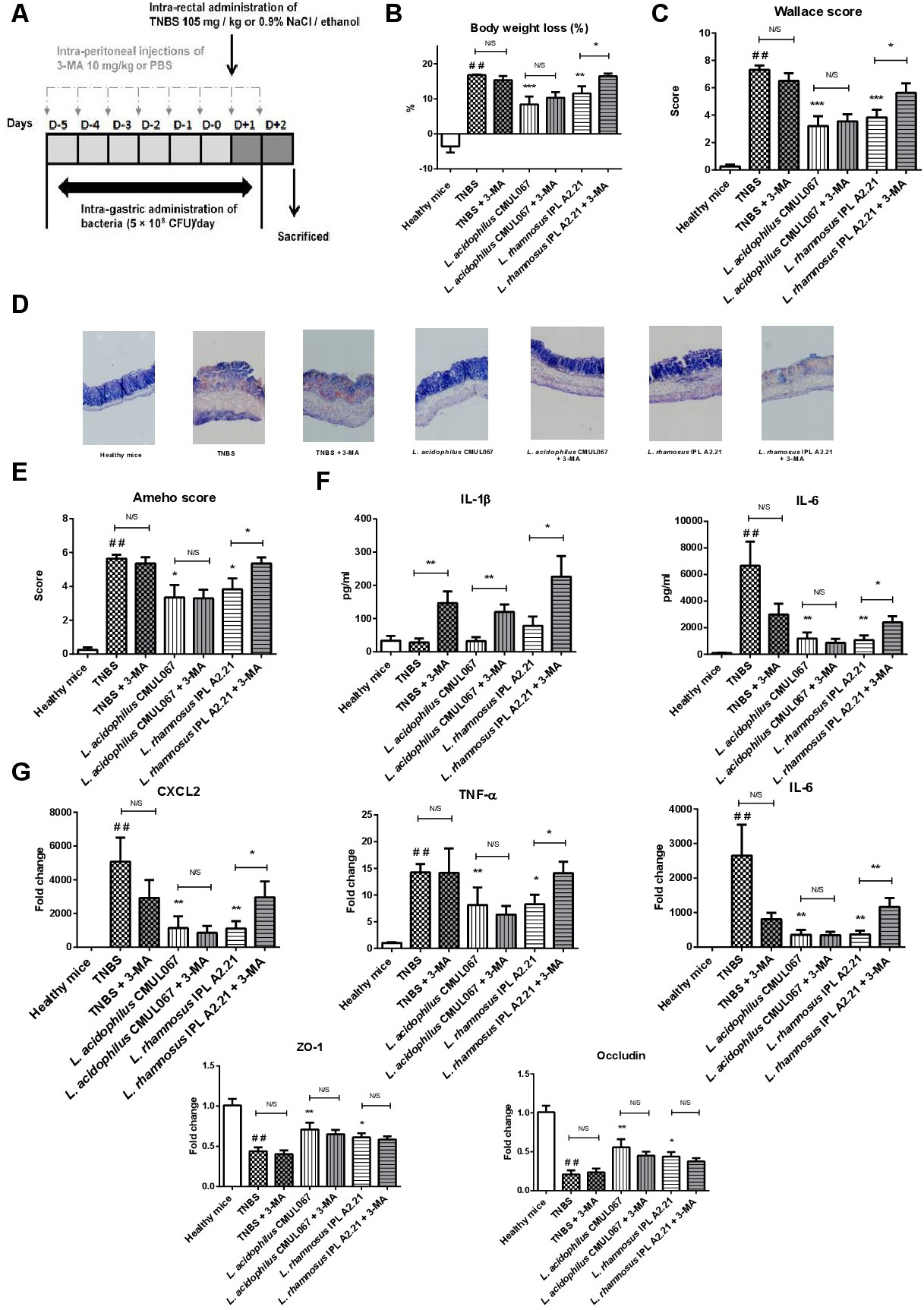
Fig. 3. In vivo inhibition of autophagy limited the capacity of L. rhamnosus A2.21 to rescue mice from acute TNBS-induced colitis. (A) Experimental protocol used for the mouse model of acute colitis in BALB/c mice was induced by intrarectal administration of TNBS (105 mg/Kg), intragastric administration of bacteria and IP injection of 3-MA. (B) Body weight loss (as a percentage of the initial weight) (C) Macroscopic evaluation of colonic inflammation (Wallace score) (D) Representative histological sections (stained by May Grünwald Giemsa, 100X magnification) of mice treated (TNBS) or not (Healthy mice) with TNBS, and orally administrated or not with the selected strains and treated or not by IP injection of 3-MA (E) Histologic evaluation of colonic inflammation (Ameho score) (F) Plasmatic IL-6 and IL-1β concentrations measured by ELISA, two days after colitis induction (G) Gene expression of cxcl2, il6, tnfa, zo1, and occludin from colonic samples. Values are expressed as the relative mRNA levels of samples compared with colons from healthy mice. The data represent the mean values of each group (n=10) ± SEM. * and # refer to the comparison of bacteria-treated groups versus TNBS control group or TNBS group versus healthy mice, respectively; *p<0.05, **or## p<0.01, ***p<0.001.